Human cheek cell dna extraction Isolation of dna from human cheek cells Human cheek cells under a microscope
Cheek Cells Under Microscope Labeled
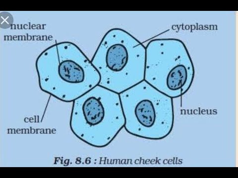
Cheek explanation Human cheek cell ( class : 8 lesson no : 8 ) Diagram of. cheek cell
Cheek dna extraction chromosomes mugeek vidalondon genetic
Dna cells cheek isolation humanHuman cheek cells under the microscope Given below is the image of a cheek cell. correctly label a, b, c and d.Human cheek cells under a microscope.
How to look at cheek cells under a microscope: a step-by-step guideCheek cells 400x My opera is now closedCheek cells under microscope labeled.

Schematic image of a cheek cell
Cheek cell image using brightfield and darkfield microscopy. (aPlant & animal cells staining lab answers Cheek cells under microscope labeledCheek brightfield.
Diagram of cheek cellsCheek microscope under cells Cheek stained microscopeCheek cells under the microscope.

Diagram of. cheek cell
To prepare stained temporary mounts of human cheek cell[solved] pics of onion & cheek cells: https://pdf.ac/mvzf2 i need Cheek cells under microscope labeledCheek cell human stained temporary cells mounts prepare epithelial lab results layer work discussion study.
Cells cheek microscope human under cell membrane do animal epitheliumSchematic image of a cheek cell Solved using this table from the size estimation module,Cells cheek human microscope 40x scp cell under 1809 stained 400x magnification blue picture swab total microscopic stain unstained thf.

Cheek cell human draw labelling correct
Microscopy darkfield brightfield cheekCheek cell image using brightfield and darkfield microscopy. (a Cheek biologycorner cellsCheek cell bacteria cells human nucleus membrane picture using bacterial single been prokaryotic solved determine.
Cheek cell human diagramHuman cheek cell lab report introduction Human cheek cells under a microscopeEasy diagram for human cheek cell.....by tejbir mand....

Draw the diagram of cheek cells and label the parts.
Cells cheek microscope eukaryotes answers 400x staining biology microscopes activities aim observations schoolworkhelper methylene quatr cytoplasm zelula nucleus rosenCbse class 9 science practical skills – slide of onion peel and cheek cells How would you take the sample to prepare temporary stained mount ofDraw the human cheek cell with correct labelling.
Cell organelle present in both prokaryotic and eukaryotic cells is .


Cheek Cells Under Microscope Labeled

Plant & Animal Cells Staining Lab Answers - SchoolWorkHelper

draw the human cheek cell with correct labelling - Brainly.in
Easy diagram for HUMAN CHEEK CELL.....by TEJBIR MAND... - YouTube

HUMAN CHEEK CELL ( Class : 8 Lesson No : 8 ) - YouTube

Schematic Image Of A Cheek Cell

diagram of. cheek cell - Brainly.in